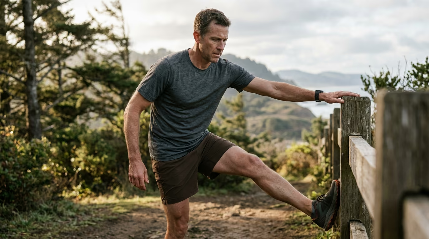

Six distinct lower-limb profiles and tissue capacity thresholds explain how endurance athletes can identify, interpret, and manage progressive training pain.





Why does your Achilles tendon ache when you step out of bed, or why does the outside of your knee throb after forty minutes of running? You type those exact questions into search engines, hoping for a simple diagnosis and a quick remedy. Most online searches return generic advice telling you to stretch your calves, buy different shoes, or stop training entirely.
This reference guide provides a definitive, science-backed framework for understanding endurance overuse injuries. It covers the mechanical principles of tissue capacity, the diagnostic realities across running, cycling, triathlon, and hiking, and the practical steps needed to keep your body resilient over decades of training.
An overuse injury develops when repetitive mechanical demands exceed the capacity of a specific anatomical structure to tolerate, recover from, and adapt to those demands. Unlike acute traumatic injuries that occur in a single instant, overuse conditions represent a gradual breakdown in the physiological balance between tissue breakdown and biological repair.
Every endurance session applies stress to your bones, tendons, ligaments, cartilage, and muscles. In response to this mechanical stimulus, your body initiates a remodeling process. When training stress is followed by adequate recovery and nutrition, tissues rebuild stronger. When the rate of micro-damage outpaces cellular synthesis, the structural integrity of the tissue declines.
Sports scientists distinguish between external load and internal load. External load refers to the physical work you perform, such as mileage, elevation gain, wattage, speed, and repetitions. Internal load reflects the physiological and psychological stress that your body experiences during that work, measured through heart rate, perceived exertion, blood lactate, and fatigue.
The International Olympic Committee consensus on training load emphasizes that injury risk stems from the complex interaction between external workload and internal physiological strain. A weekly running volume of forty miles might represent an appropriate stimulus for an athlete with years of consistent training. That same volume can overwhelm an athlete returning from a long layoff or managing severe occupational stress.
Tissue capacity is not a static number. It fluctuates based on training history, sleep quality, energy intake, systemic health, and age. Different tissues also adapt at vastly different rates:
When athletes increase training demands faster than these connective structures can adapt, the weakest point in the kinetic chain begins to signal distress. Overuse is not a sign that endurance sport is inherently destructive. It is proof of a temporary mismatch between the load applied and your current physiological tolerance.
Endurance athletes frequently struggle to distinguish between normal training soreness and the early stages of a clinical overuse injury. Pain is a vital neurological signal, but pain intensity alone does not indicate the structural severity of a condition. A minor tendon irritation can feel intensely sharp during early morning movement, while a high-grade bone stress injury might present as a dull, tolerable ache.
To interpret your physical symptoms accurately, you must track multiple clinical dimensions rather than focusing solely on pain intensity:
Diffuse discomfort spread across a broad muscle belly often points to benign delayed onset muscle soreness or generalized fatigue. In contrast, pinpoint tenderness over a bony prominence or a specific tendon segment suggests localized structural overload. If you can cover the painful area with the tip of a single finger, the tissue requires careful monitoring and potential clinical assessment.
Pay close attention to when symptoms appear relative to your training sessions:
Assess how the symptom influences non-sport activities. If an ache in your lower leg alters how you walk down the stairs, carry groceries, or stand at your desk, the injury has progressed beyond mild exercise-induced irritation. Compensatory movement patterns can quickly introduce secondary overuse issues in adjacent joints.
You should pause impact training and seek professional medical evaluation from a sports physician or physical therapist if you experience any of the following signs:
Reviewing evidence-based injury prevention resources can help you establish structured protocols before minor symptoms turn into severe interruptions.
Running and hiking impose substantial repetitive impact and eccentric braking forces on the lower extremities. Epidemiological research published in sports medicine reviews shows that annual injury incidence among recreational runners ranges from 37% to 56%. Between 50% and 75% of these problems are classified as overuse injuries.
Systematic reviews identify Achilles tendinopathy as one of the most common running injuries, accounting for roughly 10.3% of injury incidence and 6.6% of injury prevalence. The condition is characterized by localized pain and morning stiffness in the main tendon body or at its insertion on the calcaneus.
Tendinopathy involves cellular disorganization, increased neurovascular ingrowth, and altered collagen structure rather than simple inflammation. Treating a tendon solely with anti-inflammatory medication or complete rest does not restore its capacity. Tendons require carefully calibrated mechanical tension to stimulate collagen alignment and remodeling.
Medial tibial stress syndrome, commonly referred to as shin splints, accounts for approximately 9.4% of running injury incidence. Athletes feel diffuse tenderness along the posteromedial border of the distal tibia, typically spanning five centimeters or more.
The condition arises from traction of the deep lower-leg musculature and repetitive bending strain on the tibial cortex. While common among newer runners, shin pain must be monitored closely to ensure it does not escalate into a true tibial stress fracture.
Bone stress injuries represent a continuous spectrum ranging from microscopic bone strain and periosteal edema to full cortical stress fractures. Research published in the British Journal of Sports Medicine emphasizes that bone adapts to mechanical stress through remodeling, but rapid volume increases can trigger localized microdamage faster than osteoblasts can deposit new bone matrix.
Common sites for endurance athletes include the tibia, metatarsals, fibula, and femoral neck. High-risk locations, such as the anterior tibial cortex and femoral neck, carry a higher risk of non-union and require strict clinical supervision. Any localized, deep bony ache that worsens with every step demands immediate diagnostic imaging, typically via magnetic resonance imaging.
Patellofemoral pain syndrome represents anterior knee pain centered around or behind the patella, accounting for up to 16.7% of running injury prevalence. Symptoms are aggravated by compressive loading activities, including downhill running, stair descent, and prolonged deep squatting.
Modern clinical consensus rejects the idea that patellofemoral pain is caused purely by poor patellar tracking. Instead, it reflects joint overload driven by quadriceps weakness, hip abductor deficits, altered foot mechanics, and sudden training volume spikes. Comprehensive rehabilitation involves targeted hip and knee strengthening paired with temporary load management.
Iliotibial band syndrome presents as sharp or burning pain over the lateral femoral epicondyle on the outside of the knee. The discomfort typically emerges at a predictable distance during a run, especially during downhill descents or slow jogging where the knee stays near thirty degrees of flexion.
The underlying mechanism involves compression of the innervated fat pad beneath the iliotibial band rather than structural friction. Managing the condition requires strengthening the gluteus medius and maximus, modifying running cadence, and temporarily eliminating aggravating terrain.
Plantar fasciitis, or plantar fasciopathy, involves pain on the plantar surface of the heel that is sharply aggravated during the first steps in the morning. Systematic reviews show it accounts for roughly 6.1% to 7.9% of running injuries. Contributors include restricted ankle dorsiflexion, reduced calf capacity, unsupportive footwear, and rapid increases in walking or standing volume.
Hikers and ultrarunners encounter unique mechanical stressors compared to road runners. Carrying a heavy backpack shifts the body's center of mass and amplifies compressive loads across the knees, ankles, and spine.
Sustained downhill descents generate continuous eccentric contractions in the quadriceps and patellar tendon, inducing profound muscle damage and joint strain. Navigating technical, uneven trails demands constant lateral ankle stabilization, increasing the risk of peroneal tendon irritation and posterior tibial tendon fatigue.
Cycling is a low-impact sport, but its repetitive nature makes cyclists susceptible to distinct overuse conditions. A cyclist turning pedals at 90 revolutions per minute completes 5,400 pedal strokes every hour. Any mechanical inefficiency, muscular imbalance, or load spike is repeated thousands of times per ride.
A systematic review on cycling injuries indicates that patellofemoral pain is the leading non-traumatic complaint among road cyclists. However, research reveals that many traditional bike-fit measurements do not correlate reliably with overuse symptoms. Load variables, such as total weekly hours, high-gear climbing, and low-cadence grinding, often play a far more decisive role in symptom onset than saddle position alone.
Anterior knee pain in cyclists often stems from excessive patellofemoral compression. Riding at low cadences against high resistance, pushing oversized gears up steep gradients, or using an excessively low saddle height increases compressive forces behind the patella.
Posterior knee pain, involving the distal hamstrings or popliteus tendon, frequently develops from an overextended saddle height, excessive cleat float, or sudden increases in high-power sprint intervals.
Maintaining an aerodynamic or forward-leaning posture places sustained isometric demands on the spinal stabilizers, neck extensors, and upper extremities:
Addressing these issues requires a balanced approach combining bike setup adjustments, core stability, and targeted recovery and mobility strategies.
Triathletes face a unique physiological challenge because they accumulate cumulative musculoskeletal stress across three distinct disciplines. A triathlete might maintain a conservative running mileage, yet develop lower-extremity breakdown because their running sessions are performed on fatigued legs immediately following heavy cycling blocks.
Swimming places high repetitive demands on the rotator cuff and subacromial structures. Cycling forces the hip flexors and lower back into sustained flexion. Running then demands impact tolerance, eccentric control, and upright postural stability.
Transition sessions, commonly known as brick workouts, alter neuromuscular recruitment and running mechanics. When triathletes add brick sessions without adjusting their overall weekly volume, the combined internal fatigue degrades joint mechanics and elevates tissue strain.
For years, I capped my mid ride fueling at around sixty grams of carbs per hour, convinced that taking in more would wreck my stomach. Then I read a series of recent studies on gut training and higher oxidation limits for endurance athletes. I spent a three month base phase gradually increasing my intake up to ninety grams using a mix of glucose and fructose. The difference during my next Gran Fondo was staggering. I had a late race surge that I had never experienced before, completely avoiding the usual energy crash.
Proper fueling protects against systemic fatigue, helping you maintain coordinated biomechanics when training volume peaks. Exploring structured fueling and hydration approaches provides the energetic support needed to prevent fatigue-related tissue breakdown.
Most overuse injuries are not caused by bad luck or faulty anatomy. They are driven by predictable errors in training distribution, progression, and physiological recovery. Understanding these variables allows you to build sustainable fitness while protecting vulnerable tissues.
The conventional guideline that runners should never increase weekly mileage by more than 10% is widely cited, but sports medicine research offers little empirical support for it as an absolute rule. A systematic review published in the British Journal of Sports Medicine found limited evidence that sudden changes in running volume alone predict injury when examined in isolation.
Injury risk is shaped by the interaction of volume, intensity, surface, and cadence. An athlete might increase weekly distance by only 5%, but if they add high-intensity track intervals and hill sprints in the same week, the mechanical load on their Achilles tendons and shin bones spikes dramatically. Training progression must account for the total mechanical stress across all variables.
The acute-to-chronic workload ratio compares the training load performed during the current week to the rolling average of the preceding four weeks. The model suggests that maintaining a ratio between 0.8 and 1.3 keeps athletes in a safe zone, while ratios approaching or exceeding 1.5 increase injury risk.
While this concept provides a helpful mental model for avoiding sudden training spikes, it has significant methodological limitations:
Performing the same workout at the same moderate intensity day after day creates high cumulative tissue strain. When connective tissues are subjected to identical mechanical vectors without variation in terrain, pace, or movement patterns, specific micro-zones within the tendon or bone experience continuous micro-trauma.
Periodized plans that alternate easy aerobic days, structured speed workouts, non-impact cross-training, and dedicated rest allow tissues to recover through varied loading patterns.
Low Energy Availability occurs when an athlete's dietary energy intake is insufficient to support physiological functions after subtracting the energy expended in exercise. In both male and female athletes, prolonged low energy availability impairs bone mineral density, reduces protein synthesis, suppresses sex hormones, and elevates bone stress injury risk.
Bone health depends on adequate caloric intake, optimal calcium and vitamin D levels, and balanced hormonal function. Athletes who severely restrict calories while sustaining high endurance volume undermine their body's capacity to repair skeletal microdamage, creating a direct path toward stress fractures.
Epidemiological studies consistently identify previous injury as the single strongest risk factor for a future overuse injury. A prior injury often leaves residual deficits in local muscular strength, reduced neuromuscular coordination, altered movement mechanics, or compromised tissue architecture. Rushing back to full training before achieving complete structural rehabilitation almost guarantees recurrence.
Integrating evidence-based concepts from training and performance guides helps ensure that your training load builds progressively upon established baselines.
Endurance athletes over thirty-five can achieve exceptional levels of aerobic fitness, endurance capacity, and competitive success. However, biological aging introduces physiological shifts in tissue compliance, cellular repair rates, and hormonal profiles that require thoughtful training adjustments.
As the body ages, collagen synthesis slows and connective tissues lose water content. Tendons and ligaments develop more advanced glycation end-products, altering their natural elasticity.
An older tendon cannot absorb and release elastic energy as quickly as a younger tendon, making it more vulnerable to sudden spikes in plyometric or sprint loading. Older athletes require longer adaptation periods when introducing speed work, steep hill repeats, or sudden changes in footwear drop.
Age-related muscle loss, known as sarcopenia, preferentially affects type II fast-twitch muscle fibers. These fibers provide the eccentric force production required to brake the body during downhill running and stabilize the pelvis during high-cadence cycling.
When stabilizing muscles fatigue prematurely, impact forces are transferred directly to bones, articular cartilage, and passive connective tissues. Incorporating heavy, slow resistance training two days per week preserves fast-twitch motor units and enhances structural capacity.
Both men and women experience endocrine changes with age. Declining estrogen levels during perimenopause and postmenopause accelerate bone resorption, making female masters athletes particularly susceptible to bone stress injuries and osteopenia.
In men, gradual reductions in testosterone and growth hormone can slow systemic recovery times and reduce muscle protein synthesis. Monitoring bone mineral density through periodic dual-energy X-ray absorptiometry scans provides essential data for older endurance athletes.
A twenty-five-year-old athlete may fully recover from an exhaustive interval session in 36 to 48 hours. A fifty-year-old athlete might require 72 hours to achieve the same muscular and cellular recovery. Attempting to maintain rigid, multi-day high-intensity training schedules without extended recovery windows leads directly to chronic overtraining and overuse pathology.
Understanding the physiological nuances detailed in healthy aging resources allows older athletes to sustain high performance while respecting their body's evolving recovery needs.
Managing an overuse injury does not require complete immobilization. In modern sports medicine, total rest is rarely the solution for connective tissue injuries. Extended bed rest or total cessation of movement leads to rapid muscle atrophy, reduced tendon stiffness, decreased bone density, and impaired joint nutrition.
Instead, the modern standard of care emphasizes relative rest and mechanotherapy: applying progressive, appropriately dosed mechanical load to stimulate tissue healing without aggravating symptoms.
The initial step is to identify and reduce the specific aggravating movement patterns while maintaining cardiovascular fitness through non-provocative cross-training:
Once acute pain subsides, structured resistance training must be introduced to rebuild tissue capacity:
Subtle adjustments to your movement mechanics can significantly reduce tissue strain without compromising performance:
Returning to full training must follow a structured, criteria-based progression rather than an arbitrary timeline. Progress from low-load aerobic sessions on flat terrain to moderate endurance volume before reintroducing high-intensity intervals, hill climbing, or competitive events.
You cannot manage what you do not measure. To catch overuse injuries before they develop into chronic conditions, you must systematically track both external workloads and internal recovery metrics.
Every morning, score the following variables on a simple 1 to 5 scale before getting out of bed:
If your cumulative score drops significantly below your baseline for two consecutive days, adjust your scheduled workout from high-intensity intervals to an easy aerobic recovery session or a rest day.
Calculate your training load for every workout by multiplying the session duration in minutes by your Rating of Perceived Exertion on a 1 to 10 scale. A 60-minute tempo run rated at an RPE of 7 equals a training load of 420 units.
Tracking this metric ensures that you monitor the combined mechanical and physiological demands of your training rather than relying on mileage alone.
Athletes managing early-stage tendon or bone sensitivity should establish a standardized morning palpation test. Firmly squeeze the affected tendon or press the bony surface each morning and rate the localized tenderness on a 0 to 10 scale.
If morning tenderness jumps by more than two points following a workout, the previous day's training load exceeded the tissue's current tolerance. Reduce the load for the next 48 hours until tenderness returns to baseline.
Reviewing practical recovery methodologies from recovery and adaptation resources will help you establish consistent daily monitoring habits.
To apply the science of load management and tissue capacity to your own endurance routine, use this practical checklist over the coming week:

Follow ReEndure for practical insights on endurance training, recovery, nutrition and healthy aging. Stay connected for new articles, research led guidance and ideas to help you perform better for longer.


Read practical ideas on endurance training, recovery, nutrition and healthy aging to keep progressing for years to come.
Read the Blog