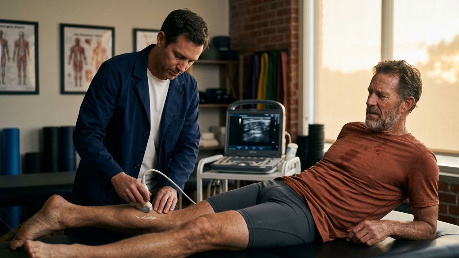

A recent biomechanics study found ultrasound temporarily reduces deep calf stiffness. Learn why masters endurance athletes still need progressive loading.





On September 1, 2026, the Journal of Biomechanics published a new paper examining therapeutic ultrasound. The study appeared in Volume 206 of the publication. It was previously made available online on August 6, 2026. Researchers from Doshisha University’s Graduate School of Health and Sports Science conducted the experiment.
They wanted to determine if ultrasound could alter passive stiffness in deep lower leg tissues. The researchers tested the calf muscles of 20 healthy adults. This paper carries the academic identifier 10.1016/j.jbiomech.2026.113502. The findings offer a specific look at how deep muscle tissue responds to noninvasive treatment.
The primary conclusion centers on a distinct physical difference between two parts of the calf complex. The ultrasound intervention significantly reduced shear-wave velocity in the soleus muscle. This metric serves as an imaging-derived indicator of passive stiffness. The untreated opposite leg showed no significant change during the experiment.
The same study found no significant change in the more superficial medial gastrocnemius muscle. The anatomical structure of the lower leg helps explain this contrasting result. The report states that the soleus is approximately 2.44 centimeters beneath the skin. The gastrocnemius rests much closer to the surface at approximately 0.53 centimeters.
This physical difference clarifies why the researchers focused on deeper muscle penetration. Tomohiro Umeda was one of the researchers involved in the project. Umeda said the findings suggest ultrasound may have the potential to non-invasively decrease passive stiffness in deep muscles such as the soleus.
The research team designed a precise laboratory protocol for the healthy adult participants. They treated one randomly selected leg per person with continuous ultrasound. This treatment lasted for exactly 10 minutes. The equipment was calibrated to 1 megahertz and 2.0 W/cm2.
The opposite leg served as an untreated control to establish a reliable baseline. Participants had their muscle stiffness assessed immediately before the intervention. They were tested again immediately after the treatment concluded.
The researchers utilized shear-wave elastography to capture these physiological changes. Participants who began with stiffer soleus muscles tended to experience larger reductions in stiffness. The experiment measured an immediate response rather than a permanent structural adaptation.
The study did not test whether ultrasound improved actual athletic performance. It also did not measure if the therapy shortened recovery times or prevented future injuries. The outcome simply established a temporary mechanical change in a controlled setting.
The calf is not a single uniform muscle group. It consists of multiple distinct structures that handle different physical tasks. The gastrocnemius is the larger visible muscle that crosses both the knee and ankle joints. The soleus sits deeper and only crosses the ankle joint.
This deeper muscle is often referred to as the workhorse of distance running. It absorbs massive amounts of force with every single foot strike. Endurance athletes place extreme, repetitive demands on this specific deep tissue. The Doshisha University study provides valuable insight by isolating the soleus response.
The differing depths of these muscles require different therapeutic approaches. The superficial medial gastrocnemius is easily accessible to standard manual therapy. The deeper soleus is historically harder to target with noninvasive physical treatments. The 2.44 centimeter depth of the soleus presents a unique clinical challenge.
The ultrasound waves in this experiment successfully reached and influenced this deeper layer. The fact that the 0.53 centimeter deep gastrocnemius showed no significant change is intriguing. It suggests that specific ultrasound frequencies might bypass superficial tissues to affect deeper structures.
Ambitious athletes must remain critical when reading early sports science headlines. This specific research represents a single laboratory finding in a controlled environment. The 20 participants were entirely healthy adults without chronic lower leg injuries. The study did not include endurance athletes struggling with persistent calf tightness.
It also excluded individuals actively recovering from acute calf strains. We cannot assume these findings will generalize accurately to a symptomatic athletic population. The researchers acknowledged these limitations when discussing the clinical impact of their work.
Furthermore, the recorded outcome was strictly an immediate post-treatment measurement. The study design did not establish how long the reduction in soleus stiffness lasted. A treatment that reduces stiffness for ten minutes has limited practical utility. A reduction that lasts for several hours could be incredibly valuable for rehabilitation.
Reduced passive stiffness is also not synonymous with improved athletic performance. It does not automatically guarantee greater running economy or a lower future injury risk. The researchers framed injury prevention as a potential future application rather than a proven outcome.
Modern sports medicine increasingly utilizes advanced imaging as an assessment tool. Ultrasound technology now allows clinicians to quantify tissue stiffness and viscoelastic behavior. This gives practitioners a way to monitor tissue health objectively. Dynamic ultrasound is also being used to assess calf and Achilles architecture during movement.
It tracks tendon elongation and tendon strain during controlled loading exercises. These precise imaging methods can help clinicians individualize rehabilitation protocols. They provide actual data on how tissues respond to stress.
This objective measurement approach aligns with modern clinical best practices. A qualitative study involving 20 expert sports clinicians recently examined lower leg rehabilitation. These experts found that calf rehabilitation is generally individualized. They base treatment on athlete characteristics, injury features, and specific sport demands.
Our injury prevention strategies for endurance athletes rely on this personalized approach. Clinicians avoid treating a single stiffness measurement as a stand-alone diagnosis.
Endurance athletes must adapt their training as their tissues age. Hitting my forties brought a harsh reality check. The track workouts were not getting slower, but the days after them felt significantly heavier. Instead of forcing my old Tuesday and Thursday intensity schedule, I looked at the data on Masters athletes and muscle protein synthesis.
I pushed my second hard session to Friday, allowing an extra forty eight hours of low intensity recovery. My total weekly volume stayed the same, but the quality of my intervals skyrocketed. Older tissues require more time to clear fatigue and rebuild structural integrity. Athletes frequently struggle with muscle recovery slowdowns after age 35.
We cannot simply copy the protocols we used in our twenties. Passive treatments like ultrasound might offer temporary relief for stiff muscles. They cannot replace the fundamental need for adequate rest and sensible scheduling.
Athletes should view therapeutic ultrasound as a possible adjunct rather than a standalone solution. A temporary reduction in soleus stiffness does not equal a cured injury. This brief window of reduced tightness might allow an athlete to move more comfortably. It could help them complete a tissue recovery routine without fighting excess restriction.
The researchers noted that future applications could involve individualized conditioning. They specifically identified the need for more research into optimal treatment parameters. The tested dose was highly specific to a controlled experiment. Athletes should avoid copying the exact laboratory settings without professional medical guidance.
The clinical experts in the qualitative study emphasized monitoring overall calf capacity. They track the muscle's response to physical loading over time. These clinicians described rehabilitation as taking place across six phases.
The available report does not list a universal exercise sequence for those phases. It also omits any generic return-to-sport threshold. This reinforces that recovery programs must remain highly personalized.
No passive modality replaces the hard work of tissue adaptation and strength building. Progressive loading forces the muscles and tendons to become more resilient. It prepares them for the high impact forces of endurance running. A recent sports-rehabilitation review offered a word of caution regarding passive treatments.
It noted that the clinical benefit of ultrasound remains unproven in randomized controlled trials. Athletes must weigh these unproven benefits against the proven results of active strength work. Athletes dealing with persistent symptoms must prioritize active capacity development.
A stiff calf might feel better immediately after a 10 minute ultrasound session. It will only stay healthy if the athlete builds adequate strength to handle their mileage. For masters athletes, the most defensible long-term strategy remains individualized training load management.
This strategy involves developing progressive calf strength and monitoring for changes in symptoms. Chasing a single optimal stiffness value is less useful than improving actual physical function.
Therapeutic ultrasound offers an interesting temporary reduction in deep calf stiffness, but progressive loading remains the only reliable path to long-term tissue resilience.

Follow ReEndure for practical insights on endurance training, recovery, nutrition and healthy aging. Stay connected for new articles, research led guidance and ideas to help you perform better for longer.




Read practical ideas on endurance training, recovery, nutrition and healthy aging to keep progressing for years to come.
Read the Blog